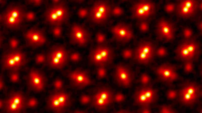
The screenshot shows an image of the PrScO3 crystal, magnified 100 million times. It was obtained using electronic ptychography
In 2018, scientists from Cornell built a powerful device that, together with a scanning technique called ptychography, set a new world record. They looked at atoms in a higher resolution than the world's best electron microscopes would allow at the time.
Despite the success the researchers have made, there was an important flaw in their approach. Their technique only worked with very thin layers of materials (no more than a few atoms thick). Any thicker sample led to the fact that the electrons "scattered", and it was impossible to somehow analyze them.
Recently, a team of experts led by David Muller broke their own record. She created an even more advanced mechanism that allows you to look at atoms in even higher resolution. And so high that only a slight haze remains in the image, which is caused by the "temperature jumps" of the atoms themselves.
The researchers' work, titled Electron Ptychography Achieves Atomic-Resolution Limits Set by Lattice Vibrations, was published in the journal Science on May 20.
“This is not just a new world record,” says Müller. - Already now we can calmly examine the atoms and identify their location. This opens up many new techniques for measuring data. What we have wanted to achieve over the years. Also, our discovery solves the problems with the study of tissues, consisting of a large number of layers of atoms. "
The new research method allowed scientists to examine individual layers of material one by one. For this, computer processing of a variety of images obtained by scattering light from the sample under study is used.
“We observe individual moving particles that our device detects, like cats observe a light from a laser pointer,” says Müller. “By tracking the behavior of moving particles in the area of layering of interference patterns, we can use a computer and calculate what the sample under study looks like at the atomic level.”
The resulting data is recreated using sophisticated algorithms, which ultimately allows you to create an image with a resolution of picometer (one trillionth of a meter).
“With such algorithms, we can eliminate almost all the causes that caused blurred images in the past. The only thing that still muddies the image a little is the mobility of atoms due to changes in temperature, says Mueller. "When we talk about temperature, we are actually talking about how much the atoms tremble."
Researchers can break their own record again if they experiment with material with heavier atoms (they are less mobile) or cool a working experimental sample. But even at zero temperature, the atoms will move, so a significant increase in the quality of the picture will not be achieved.
Electronic ptychography will allow scientists to track individual atoms in three dimensions, rather than two, as was the case in the past. It will also be possible to track those impurities that cannot be detected using classical microscopes. This will be especially useful when working with semiconductors, catalysts and quantum materials, including those used to create quantum computers, as well as when studying atoms at the boundaries of the connection of various materials. In addition, a similar research method can be used to study cells, biological tissues and even synapses in the brain.
So far, using the developments of Mueller and his colleagues is costly. It takes a lot of time and a very powerful computer to analyze all the data and compose a clear image at such a high resolution. But the researchers hope to make the method more accessible with more powerful computers and machine learning systems.
“It was like we were wearing very bad glasses all this time,” says Müller. “And now, as if for the first time, we were given a pair with high-quality diopters.”
