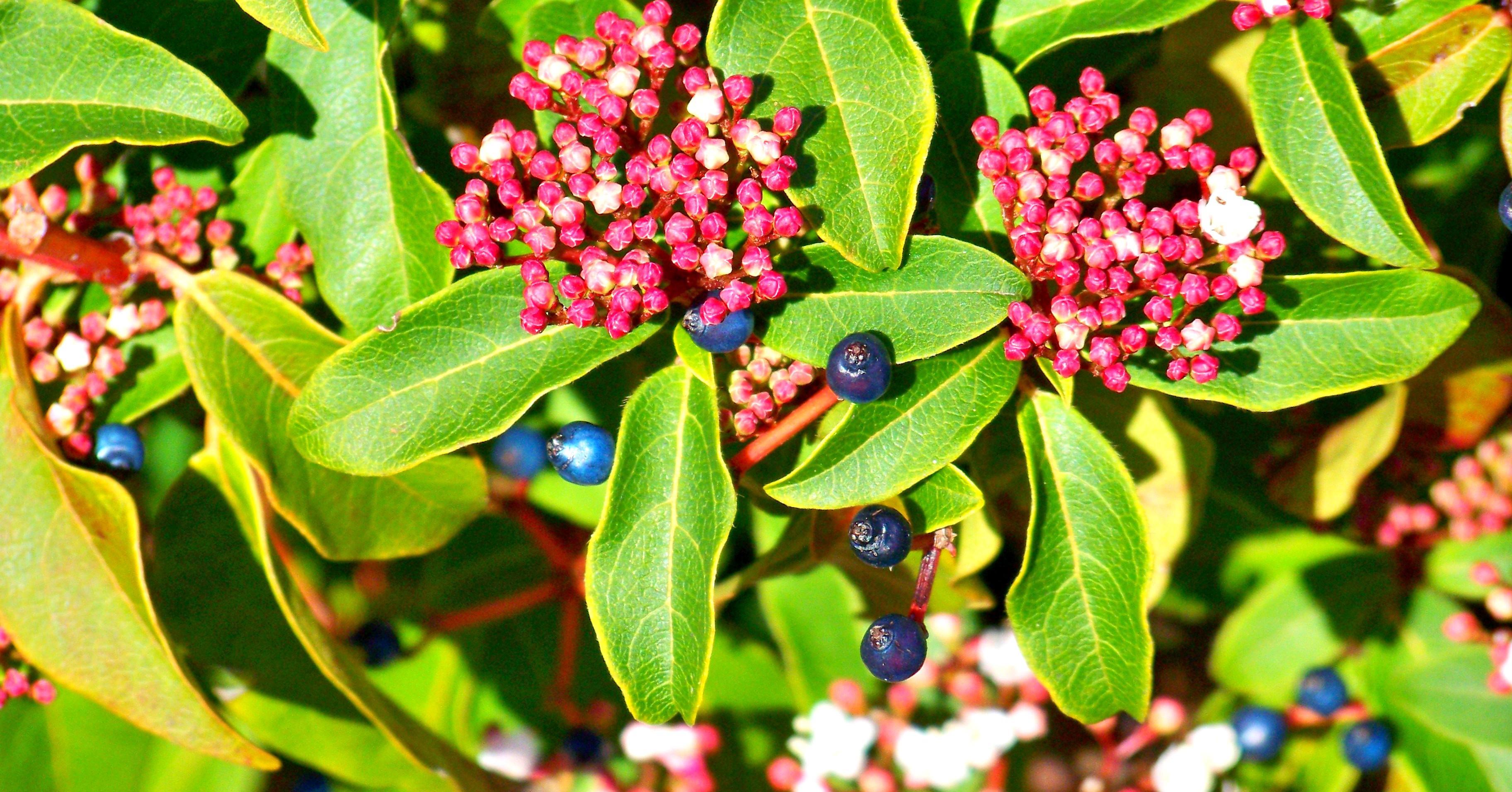
What do you associate summer with? For some, this is a period of a long-awaited vacation, for someone - a vacation, and for someone - heat, stuffiness and discomfort. If we consider summer from the point of view of gastronomy, then this is the period of vegetables, fruits and berries, which we love not only for their taste and benefits, but also for their appearance. As we know from the initial course in biology, the fruits of many plants have certain properties, the purpose of which is to attract a potential gourmet. This is an important component of the tactics of expanding the growing area. The vast majority of fruits have a bright and juicy color, indicating their goodness. The main source of this or that color in berries is pigments in the peel, but this is not the only dyeing technique. Scientists from the University of Bristol have found that laurel viburnum ( Viburnum tinus) uses lipid nanostructures in cell walls to color their berries, a previously unknown variant of structural coloration. What is so unusual about these lipid nanostructures that give the berries a dark blue color, and what is the practical application of this discovery? The report of scientists will shed light on these questions. Go.
Research results
The protagonist of this work is the laurel viburnum ( Viburnum tinus / viburnum tinus) - an evergreen bush or tree up to 6 meters in height, growing in the Mediterranean region.
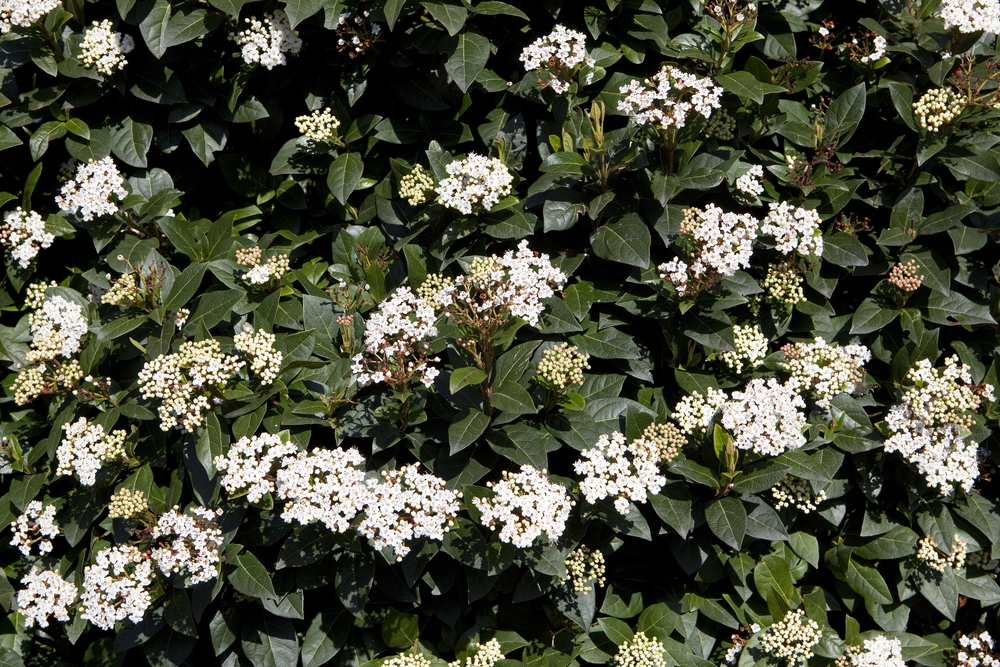
Viburnum tinus during flowering.
Viburnum tinus bears fruit several times during the year with dark blue berries, providing food for many species of birds, including the black-headed warbler (Sylvia atricapilla) and the robin (Erithacus rubecula). As with any other berry plant, birds for V. tinus viburnum are the main method of spreading seeds to new territories.
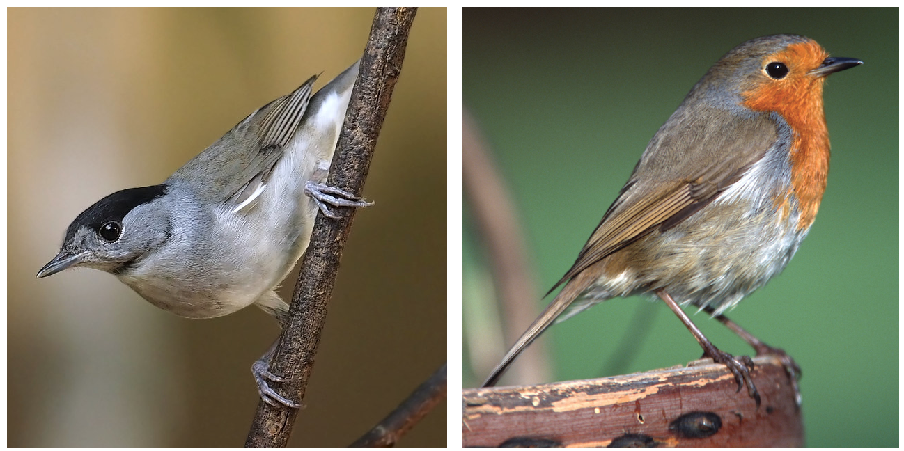
Sylvia atricapilla (left) and Erithacus rubecula (right).
At first glance, there is nothing special about this plant. A beautiful evergreen shrub that delights aesthetes among people and gourmets among birds. However, a detailed examination of the berries suggests otherwise. While other plants use different chemical compounds to color their fruits, viburnum tinus uses structural coloring. Although it was previously believed that the blue color of V. tinus berries is caused by the presence of anthocyanin pigments in their peel, this is naturally not true.
Structural colors are quite common among the fauna: butterfly wings, beetle shells, peacock feathers, etc. The color in their case is formed due to the nanoscale structural features of the surface, which cause interference with visible light.
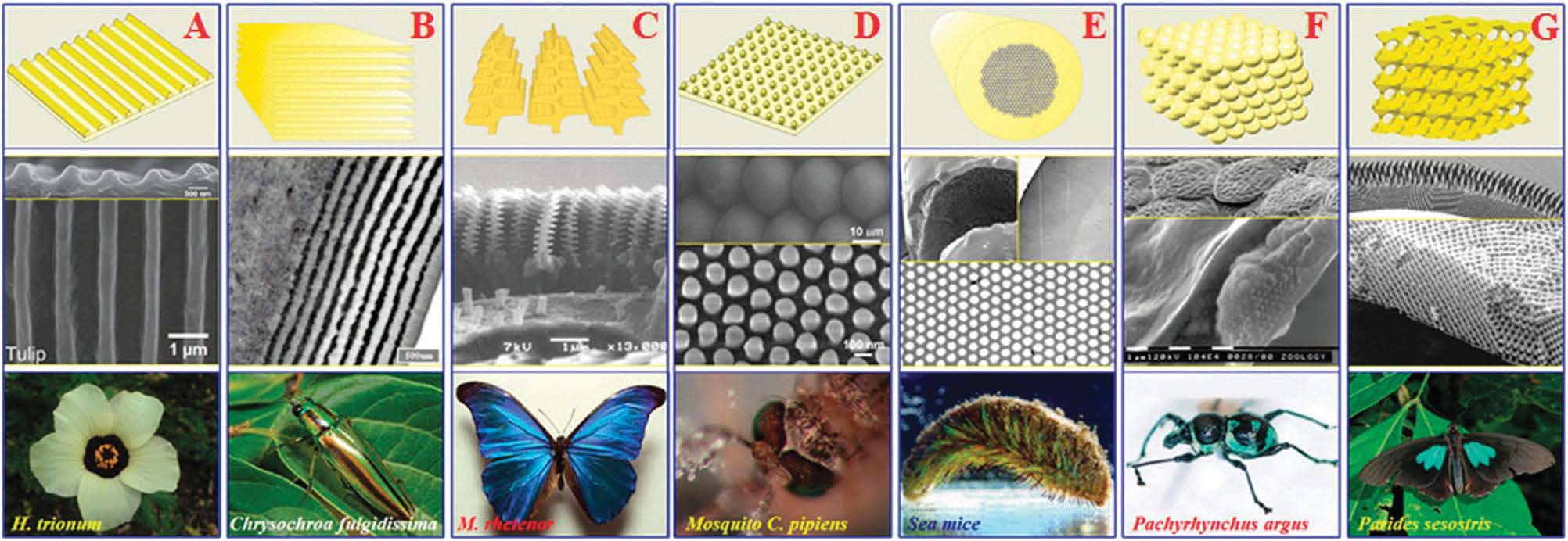
Examples of structural colors in nature: A- triple hibiscus (Hibiscus trionum); B - tamamusi beetle (Chrysochroa fulgidissima); C - butterfly of the species Morpho rhetenor; D - common mosquito (Culex pipiens); E - sea mouse (Aphrodita aculeata); F - beetle of the species Pachyrhynchus argus; G - a butterfly of the species Parides sesostris.
The peculiarity of Viburnum Tinus, which has attracted the attention of scientists, is that it not only demonstrates a new mechanism of structural staining, but is also one of the few plants capable of this.
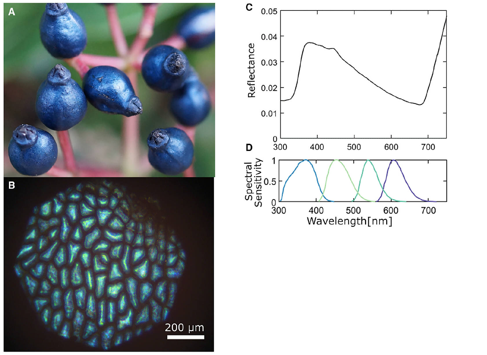
Image No. 1
Fruits of V. tinus ( 1C) reflect light directionally (giving it a metallic appearance) in the blue and ultraviolet regions of the spectrum. The polarization of the reflected light is largely retained, indicating that the coloration is structural and not pigmented, resulting from reflection from the highly structured cell wall of the outer epicarp * ( 2A ).
Epicarp * - the outer layer of the fetus.When this tissue is dissected, a dark red anthocyanin pigment is released. Light that is not reflected by the photonic structure is absorbed by the dark red pigment below it ( 2A and 3C ).
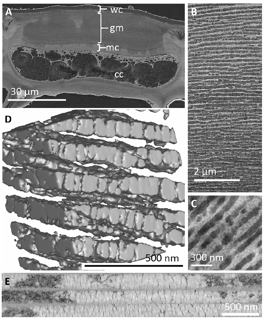
Image # 2
This absorption prevents light backscatter, increasing the visibility of blue reflection from the outer wall of the cell and thus visually improving the blue coloration.
From these observations, it can already be concluded that the color of V. tinus fruits is the result of a combination of a physical nanostructure that selectively reflects blue light waves and a base layer of blue-enhancing pigments. In other words, the interface between chemistry and physics.
To characterize the nanostructures that create the blue color in fruitsV. tinus , scientists used several electron microscopy techniques.
Scanning electron microscopy of fresh tissue ( 2A ) clearly shows the presence of a thick (10–30 µm) multilayer structure parallel to the fetal surface and embedded in the cell wall of the outermost epicarpal cells. The surface of the fruit is covered with a waxy cuticle (2–6 µm) over a layered structure. The layered architecture occupies most of the outer cell wall in the area between the cuticle and the cellulose-rich primary cell wall. The layers are 30 to 200 nm thick and cover the entire cell.
Transmission electron microscopy shows that this architecture consists of many layers of small bubbles, which differ from the matrix in electron scattering ability and refractive index.
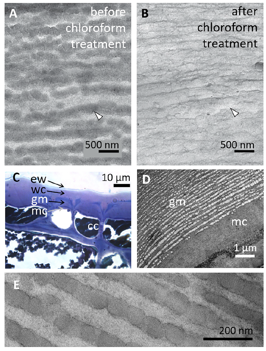
Image # 3
Scanning and transmission microscopy images show that the matrix appears to contain key components of typical plant cell walls, namely cellulose, hemicellulose and pectin. Ruthenium red ( 3D ) staining shows significant pectin content, and an electron diffraction pattern demonstrates the presence of cellulose on the characteristic diffraction rings of a natural cellulose crystal.
It should be noted that the contrasting layers are discrete and remain distinct from each other, but considerable disorder is introduced by non-parallel adjacent layers and the unevenness of their globular structure.
Imaging of the epidermal cell wall ( 2E ) shows that these globular vesicles are organized into combined layers through which the cellulose cell wall matrix remains connected by bridges and filaments ( 2B and video below).
Globular multilayer structure model (corresponds to 2D image ).
It follows from this that the globular multilayer structure of the epidermis of V. tinus fruits consists of lipids embedded in the matrix of the cell wall using various methods.
Next, ultrathin slices of the fruit epidermis were exposed to chloroform. This analysis is highly indicative since solubility in non-polar organic solvents is a clear indication of the presence of lipids.
TEM images of the same sample area before ( 3A ) and after ( 3Ba) exposure to chloroform indicates that the globular structure has been removed by treatment. In the last image, the contrast of the multilayer globular phase is reduced, while empty structures within the matrix remain visible. In comparison, exposure to water did not alter the ultrastructure or image contrast of the globular laminate, indicating that the material can only be extracted with non-polar solvents. In addition, when the scientists used imidazole-buffered osmium tetroxide (C 3 H 4 N 2 / OsO 4 ), which binds to lipids, in the process of chemical fixation , the globular layers became stained, which confirms their lipid nature.
And when ruthenium red, which binds to pectin, was used, the cell wall matrix was stained, while the globular structure was removed due to the absence of imidazole buffer.
During all staining variations applied during the study, dark outlines were observed around the globules ( 3E ). According to scientists, this may indicate the presence of a lipid membrane, theoretically necessary at the interface between hydrophobic molecules and hydrophilic polysaccharides of the secondary cell wall.
Scientists remind us that lipids are composed of a variety of molecular structures, usually classified as waxes, fats, and oils, based on their melting point.
On the surface of the epidermis of plants, waxes can easily be found, forming a water-resistant waxy cuticle. Waxes also include many molecular structures, but the predominant component is alkanes, which are virtually indigestible, i.e. have no nutritional value for birds. Fats and oils, on the other hand, are vital nutritional components, as they contain much more energy per unit volume than starch or proteins. Fats can usually be found in large quantities in seeds, i.e. deep inside the fetus.
In the case of V. tinus fruitsThe close proximity of the globular structure to both large, energy-rich seeds and the waxy outer cuticle makes the distinction between wax and fats particularly important for understanding the functional significance and origin of the structure. Therefore, it is necessary to determine whether the lipid globules are indigestible wax or nutritive fats and oils. For this, light microscopy was used.
Tissue sections of V. tinus fetus were incubated with Nile blue A (pigment), which stains the globule- rich region of the V. tinus cell wall blue or blue-violet ( 3C). This suggests that the globules are free fatty acids, and not the cutin polymer (a component of the cuticular membrane), which would turn pink or red.
In addition, the electron diffraction pattern of the multilayer globular structure shows a clear annular pattern that differs from the diagram of the cellulose cell wall with the characteristic two rings of cellulose crystals. This pattern indicates that the lipid bodies are likely crystalline and therefore are homogeneous monomeric lipids rather than polymerized molecules such as cutin.
To confirm that the observed mixed structure of cellulose matrix and layered lipid globules is responsible for the blue reflectivity of V. tinus fruits, scientists have modeled its optical response. For this, the scientists studied two mathematical models: a two-dimensional array of spheres and averaging over a set of one-dimensional two-phase multilayers.
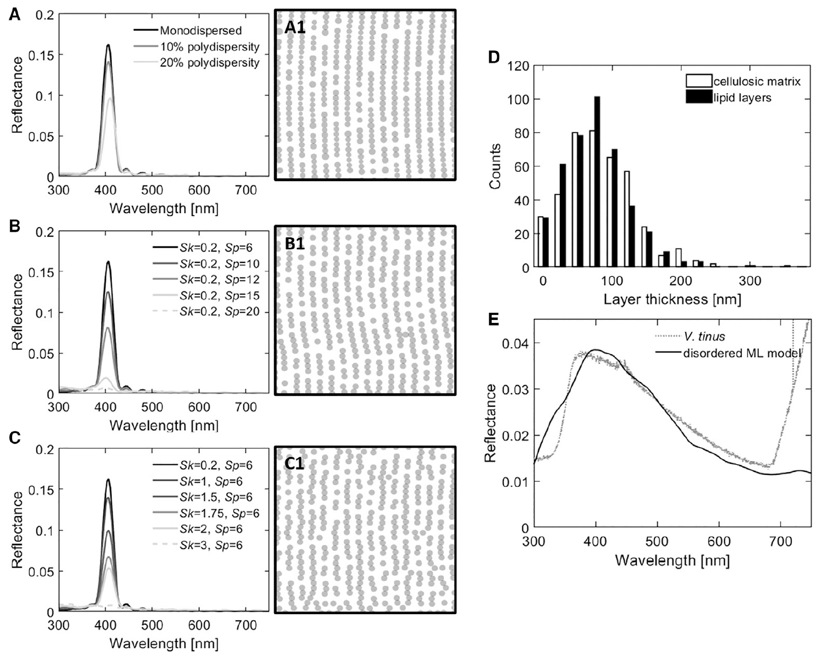
Image # 4
A reverse engineering algorithm was used to model the structure in two-dimensional space as a series of globular clusters. Schemes 4A - 4C correspond to the adjacent modeled reflectance spectrum. This algorithm allows different types of disorder to be independently introduced into the globular multilayer by adjusting the size and structure, i.e. Fourier transform of the position of particles.
In the process of modeling, the following were studied: the optical response of layered lipid globules with varying degrees of variation in the globule diameter ( 4A); disorder in the angles between adjacent globules (parameter Sp, 4B ) and disorder at the average distance between adjacent globules (parameter Sk, 4C ).
The introduction of different types of disorder ( 4A - 4C ) always had the same effect on the optical response of the globular multilayer, namely, a decrease in the peak intensity.
Thus, instead of considering each disorder element separately, the structure and material composition of the V. tinus cell wall were approximated by disordered one-dimensional multilayers with refractive indices corresponding to cellulose (n = 1.55) and typical plant lipid (n = 1.47). The thickness distribution of both materials is shown in4D . And the reflectivity, modeled using averages over 1D layers, is shown in Figure 4E .
Introducing the disorder observed in the cross section measurements into the coherent ordered reflector model broadens its reflection spectrum.
If the model allowed scientists to understand exactly how V. tinus berries get their color, then modeling does not cover the need for such a mechanism.
The greatest interspecific interaction in V. tinus is associated with birds feeding on the berries of this amazing plant. Comparison with the spectral sensitivity of the tit ( 1D) shows that the color of berries is within the range visually significant for birds of this species.
The berries, of course, do not float in the air, but are attached to the branches on which the leaves grow - a visual background. To a large extent, the background is green due to the dominant chlorophyll pigment in the leaves. Chlorophyll has a broad spectral characteristic, with a peak at 550 nm and little reflectivity below 500 nm, which gives the color of V. tinus fruits a chromatic contrast with foliage. In other words, the berries look even more noticeable against the background of such leaves.
Considering that visual signals for birds are often of priority, the lipid structural coloration of V. tinus berries can serve as a strong visual signal for hungry birds.
Considering that the color of food for birds may be the primary parameter of edibility, the color of V. tinus berries signals that they are edible and nutritious.
The relationship between the color of the fruit and its nutritional value has been studied earlier. According to some reports, the dark fruits of plants from the Brazilian region are rich in carbohydrates, while the dark fruits of plants from the Mediterranean region are rich in lipids.
Scientists believe that in the case of V. tinus, the blue color is a signal that the berries are rich in nutrient lipids, which, by the way, create this color.
Scientists call this method of signaling "honest" or "direct" when the context of the signal matches its source (blue color due to lipids - high lipid content). This signaling method is rather expensive, because the use of classical pigmentation would be easier for the plant. Nevertheless, the return that V. tinus gets in the form of attracting the attention of birds of different species, apparently, overcomes this disadvantage.
For a more detailed acquaintance with the nuances of the study, I recommend that you look into the report of scientists .
Epilogue
Color is an important component of visual information that living organisms receive about the world around them. Many animals use their color to camouflage, attract mates or scare off enemies. Some of these tactics are also present in plants, but the most important is the maintenance of interspecific communication. In the case of V. tinus, birds are the main partners of this plant, necessary for spreading seeds over long distances, which significantly increases the range of growth of V. tinus and therefore the chances of survival of the species.
The taste of the fruits of many plants depends on how much they want to attract the attention of animals of various species. Some fruits will be tasty, for example, for specific species of birds, while for everyone else they will be practically inedible. In such a complex system as interspecific communication, an important role is played by the degree of co-evolution of plant and animal species that form it.
The blue color of Viburnum laurel lies in its non-standard origin - lipid nanostructures contained in the walls of the epidermal cells of V. tinus berries . This method of staining (structural), especially due to lipids, has so far been found among plants only in V. tinus... In addition, lipid staining can tell birds about a high lipid content in berries, no matter how strange it may sound.
Honest signals, the origin of which matches their context, are quite rare in nature. The explanation for this rarity is quite simple. Imagine you own a bakery. You want to attract more customers, so you distribute flyers. Therefore, the signal has one context (we have delicious buns), but its origin is different (I don't have a bun, but something better is a drawing of a bun, i.e. the flyer is just a piece of paper). If you hand out buns, it will be an honest signaling, but much more expensive.
Previously multilayer lipid architectures like V. tinus berrieswere not seen in biomaterial. In the past, there were no such advanced tools and techniques as now, therefore many details were recorded incorrectly or were completely missed.
In the future, scientists intend to analyze other plants, which, in theory, may also have similar lipid nanostructures and, therefore, an unconventional method of fruit coloring. In addition, the scientists believe their research could help create safer food colors.
Thanks for your attention, stay curious and have a great weekend, guys! :)
A bit of advertising
Thank you for staying with us. Do you like our articles? Want to see more interesting content? Support us by placing an order or recommending to friends, cloud VPS for developers from $ 4.99 , a unique analogue of entry-level servers that we have invented for you: The Whole Truth About VPS (KVM) E5-2697 v3 (6 Cores) 10GB DDR4 480GB SSD 1Gbps from $ 19 or how to divide the server correctly? (options available with RAID1 and RAID10, up to 24 cores and up to 40GB DDR4).
Is Dell R730xd 2x cheaper in Equinix Tier IV data center in Amsterdam? Only we have 2 x Intel TetraDeca-Core Xeon 2x E5-2697v3 2.6GHz 14C 64GB DDR4 4x960GB SSD 1Gbps 100 TV from $ 199 in the Netherlands!Dell R420 - 2x E5-2430 2.2Ghz 6C 128GB DDR3 2x960GB SSD 1Gbps 100TB - From $ 99! Read about How to build the infrastructure of bldg. class with Dell R730xd E5-2650 v4 servers at a cost of 9000 euros for a penny?