Well, I hope you have already seen the world's largest laser installation 130 meters long, installed in Sarov at VNIIEF. It is intended, among other things, to study thermonuclear (!) Fusion.
This article is a transcript of a lecture by Dmitry Artemiev, senior lecturer at the Department of Laser and Biotechnical Systems at Samara University and junior researcher. research laboratory "Photonics". Dmitry gave this lecture at our Samara Boiling Point right before the introduction of the general self-isolation regime.
What is light
To complete the picture, let's start with the basics. It is known from the physics course that light is an electromagnetic wave or a stream of photons. Since one of the characteristics of electromagnetic waves is wavelength, by light (radiation) we will mean an electromagnetic wave with a length of 1 nanometer to several centimeters. Thus, our definition covers the range from X-ray to infrared radiation.
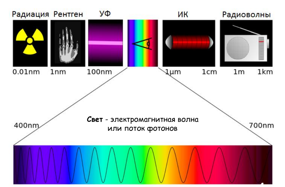
The range visible to our eyes is very small, about 300 nanometers.
If we talk about outlandish ranges, such as X-rays, then, for example, last year the creation of a free electron laser that works in the X-ray range became one of the main topics and was nominated for the Nobel Prize in Physics. Interestingly, the winner in this nomination was also associated with laser technology: the prize was awarded for the creation of ultra-short and ultra-powerful pulses. By the way, part of the research was carried out in Russia, at the Nizhny Novgorod Institute of General Physics.
How a laser differs from a conventional light bulb
The picture shows a comparison of the main characteristics. It should be especially noted that the maximum laser power is many times higher than the power of the sources used in lamps. But not every laser needs this: often a fraction of a watt, milliwatt or microwatt is enough for application to get just some specific radiation.
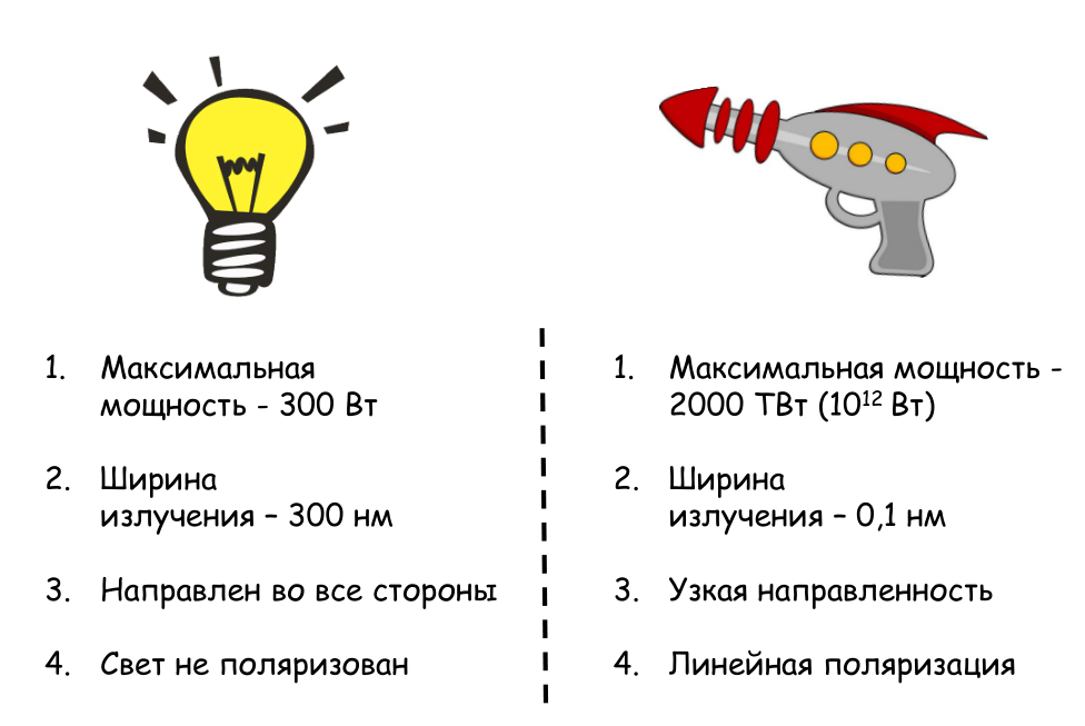
Let's remember that the width of the visible radiation range is about 400 nanometers. An incandescent lamp has approximately the same spectrum in width, so when colors are mixed, we see white light. In turn, the width of the laser range can be 0.1 nanometer. This unique property of the laser is used in some spectral studies and precise precision measurements.
If we shine a laser pointer from one side of the room to the other, we will see only a small spot on the opposite wall, demonstrating a narrow directivity of the radiation and a small divergence of the laser beam. And for fluorescent or incandescent lamps, the radiation is practically isotropic, i.e. directed in all directions.
Natural light lacks a certain directionality of the electric field vector, which means that the light is not polarized. That is, for the light of an ordinary bulb, the vector E (intensity) is directed in different directions. In the case of laser radiation, the vector E has a certain direction, the oscillations occur in the same plane. This polarization also makes laser radiation somewhat unique.
Process physics
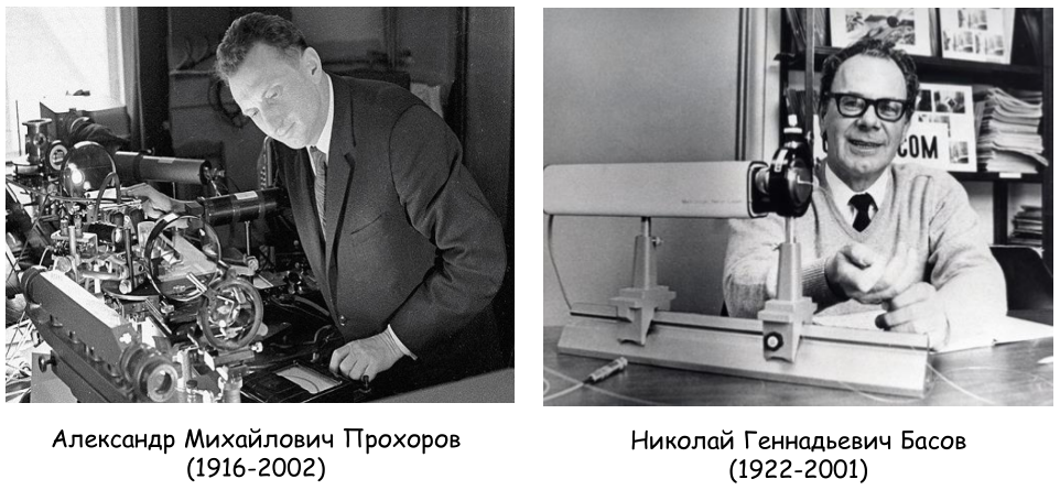
The laser was invented in the late 50s of the last century. In 1964, the American Charles Townes and Soviet scientists Alexander Mikhailovich Prokhorov and Nikolai Gennadievich Basov received the Nobel Prize for the discovery of laser radiation. Moreover, Prokhorov and Basov discovered not a laser, not an amplification of light, but an amplification of radiation in the microwave range, the so-called maser.
Laser is an abbreviation of five Latin letters: Light Amplification by Simulated Emission of Radiation. Translated from English, this means "amplification of light by stimulated radiation." Below are three diagrams. First, for radiation to occur, it is necessary for an electron or particle to pass into an excited state. For this, the particle must receive energy. After that, she will move to a higher energy level.
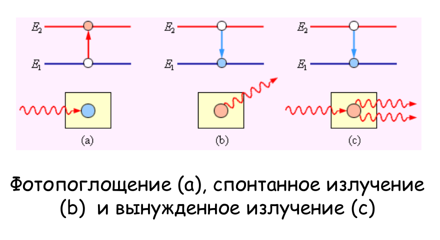
Further two scenarios are possible. If the particle randomly moves to lower energy levels, then we get spontaneous emission. However, if a particle located at the upper energy level is influenced by a certain photon, that is, to direct light of a certain wavelength at it, then a forced radiation will already occur. And the photon, born as a result of such an external influence, will be identical to the photon with which it interacted. This is how coherent radiation is obtained, in which the waves are equal to each other.
How the laser works
Here is a diagram of the first laser. This is a classic ruby laser created in 1960 by the American scientist Theodore Maiman. The device requires an active medium, in this case a ruby crystal, and two mirrors. One mirror is dull, with a reflection coefficient close to unity. The second is semitransparent, depending on the type of lasers, the reflection coefficient of it may differ by both fractions of a percent or tens of percent relative to a reflective mirror.
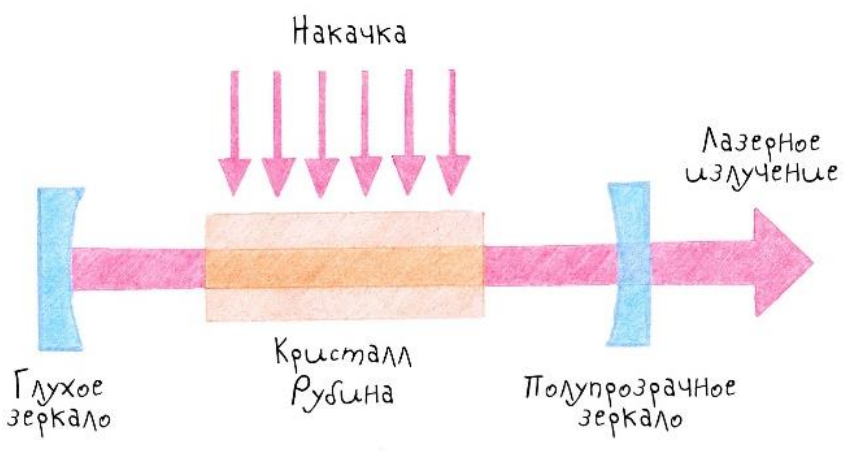
As an optical pumping for solid-state lasers, as a rule, other optical radiation is used. The first ruby crystal laser used white light lamps, which contained blue and green spectra - it is these that the ruby crystal absorbs best.
So, the classical scheme of a laser: this is an active substance (ruby), a cavity (two mirrors) and a pumping system. In other schemes, pumping can occur not only from optical radiation, but also, for example, using an electric discharge (in gas lasers). But first of all, lasers differ in the type of active medium: solid-state lasers, gas lasers, metal vapor lasers. Above, we mentioned a free electron laser; now it is being actively developed and modernized. Also, diode (semiconductor) lasers and fiber lasers, where optical fiber is used as an active medium, are now popular.
Where is laser radiation used
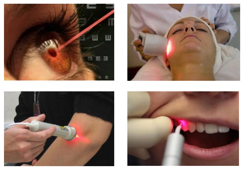
Laser radiation can be used in medicine, industry, communications, military affairs and science. The picture below shows examples of medical instruments. So, now laser scalpels for vision correction are very popular. They help correct the geometry of the lens to get rid of myopia or hyperopia, correct astigmatism, and so on. The laser is ideal for eye surgeries not only because of the very small beam size - it is also important that the exposure time with such a scalpel can be reduced to femtoseconds. Various types of radiation are used for cosmetic surgery. And in dentistry, ultraviolet radiation is used to harden dental glue, which absorbs it very well.
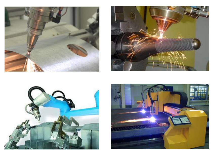
In industry, the most precise processing of steel is carried out using lasers: engraving, cutting holes with a very thin and clean edge. The properties of laser radiation are used to harden some metals. The most commonly used fiber laser in modern industry.
In the construction industry, lasers are used to determine distances or build geometry. Now laser levels are sold in all hardware stores, and they are inexpensive.

The military and hunters have been using laser sights for a long time. At the same time, the laser is rarely used for direct damage: while such devices are too bulky. For example, the American military conducted an experiment in which a laser system was installed on an aircraft. What was the whole plane for? Despite the small size of the emitter, the pumping system consumed a huge amount of electricity, and the active medium was very hot. So almost all of the plane's space was occupied by the laser power and cooling systems.
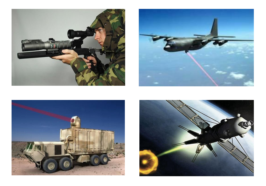
Similar systems are also being developed in our country. A couple of years ago we announced the Peresvet laser weapon. So far, the only thing known about it is that it is placed on a mobile platform, on a truck. The rest, alas, is a state secret.
Separately, it should be said about the use of lasers in scientific research. For example, scientists in Sarov use a laser in the process of thermonuclear fusion: to irradiate a target, high-power radiation is focused into a spot of minimum size.
Such lasers can take up large spaces: a thermonuclear reaction requires a serious source of radiation, the size of which can reach hundreds of meters.
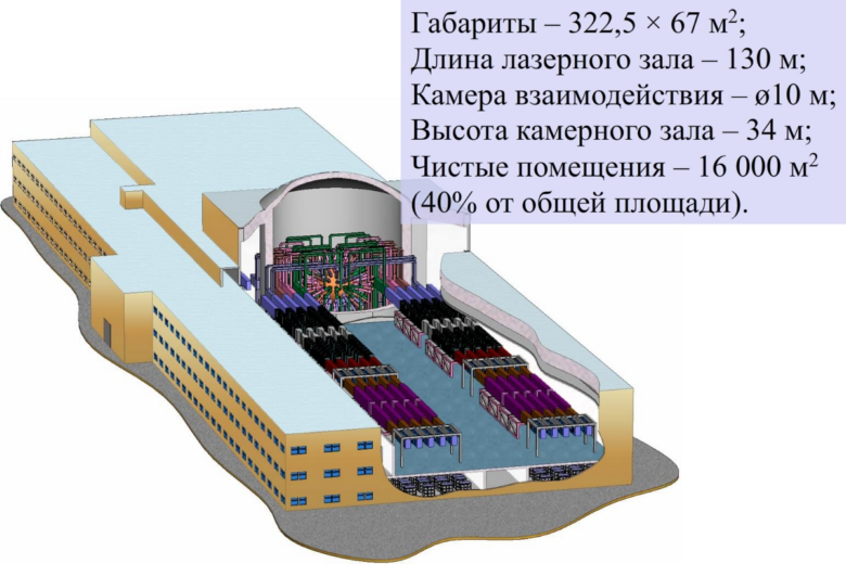
Laser facility UFL-2M in Sarov
Along with such giants, comparable in size to football stadiums, miniature lasers based on so-called nanostructures are gaining popularity lately.
Lasers are actively used in communication systems, including satellite ones. One of the most useful properties for communications workers is the propagation of radiation in an optical fiber: fiber-optic systems allow transmitting up to hundreds of gigabytes per second over great distances.
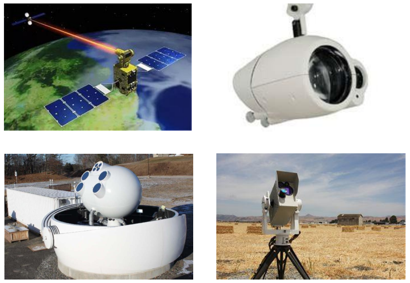
How fiber works
The principle of operation of optical fiber is based on the effect of total internal reflection. Look at the picture below: we have a stream of water, and if radiation is applied to the input, then when the stream is bent, it does not go out, but spreads inside.
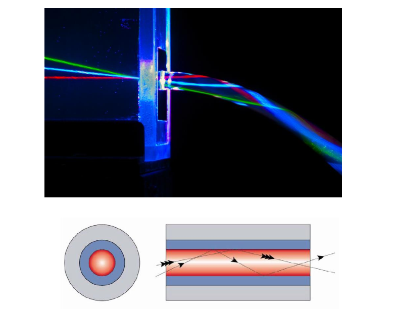
This is how radiation propagates through a medium with a higher refractive index relative to its shell. This principle allows you to transfer data over tens, hundreds and thousands of kilometers with minimal losses.
Either LEDs or laser diodes are used as a source of optical radiation. The laser diode has better performance, but it also costs more.
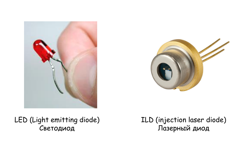
In telecommunications technology, as a rule, semiconductor lasers with a wavelength of 1.3 or 1.55 micrometers are used. These wavelengths do not fall within the absorption band of various hydroxyl groups that are present in the fiber. Thus, the signal is not absorbed or attenuated over many kilometers.
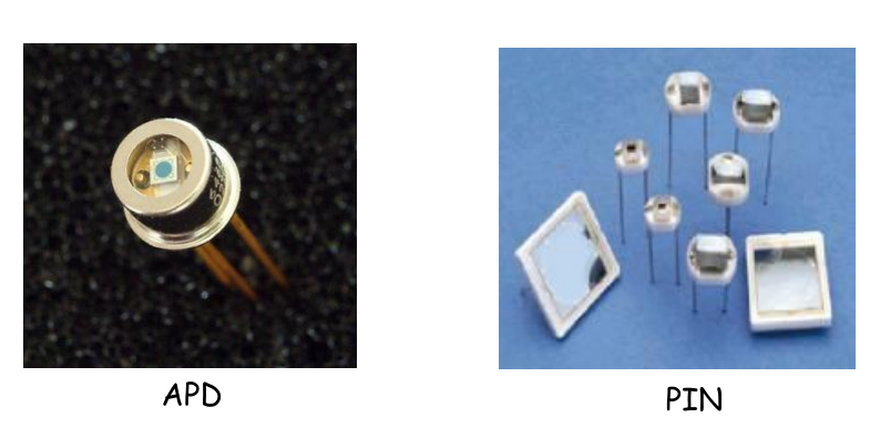
Photodiodes, PIN diodes and avalanche photodiodes can be used as detectors. They differ in sensitivity. If you want to register a very weak signal, take an avalanche photodiode. If the signal is tens to hundreds of watts, then any other types of photodiodes can be used.
Laser radiation and biological objects
When a laser beam hits a biological tissue, absorption of this radiation, as well as transmission, scattering or fluorescence, can occur. Another possible option is ablation, burning of the upper layers of tissue. In this case, the inner layers are not damaged.
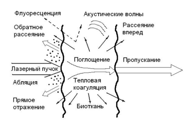
During absorption, coagulation of various particles takes place, that is, their adhesion. This effect is applied when using a laser in surgery - as a laser scalpel. Unlike a mechanical scalpel, a vessel or tissue is cut almost bloodlessly. In addition, the laser beam can be significantly thinner than the tip of a metal scalpel.
The graph below shows elements that can be found in blood vessels, in blood, in skin tissues. As we know, more than 70% of a person consists of water. Water is also present in every biological tissue. There is melanin that stains our tissue. If we get tanned in the summer, then the melanin in the skin tissues becomes significantly more. And the hemoglobin we all have can be in two states - saturated with oxygen (oxyhemoglobin) and without oxygen (deoxyhemoglobin).
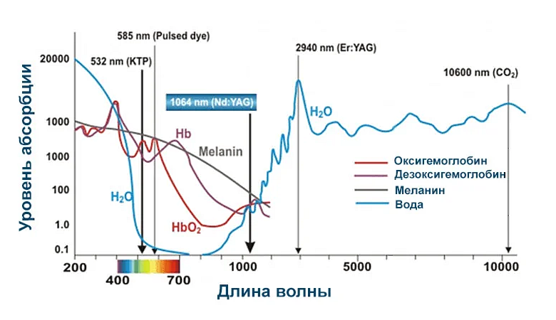
The graph shows how actively different elements absorb radiation at different wavelengths. Thus, by using a laser with a specific wavelength, we can achieve selective absorption.
Or, for example, let's take two sources of radiation with different wavelengths: one hits the absorption maximum, the other - at the minimum. With differential contrast, the concentration of certain substances can be obtained. We see that the maxima of the spectra of oxy- and deoxyhemoglobin are spaced apart. Thus, we can determine the concentration, for example, oxyhemoglobin.
This is very important when performing surgical operations. Now in any surgical department there is a device that monitors blood oxygen saturation. This sensor allows you to determine in real time what is happening with the patient's tissue in the right place.
Diagnosis, imaging, cancer treatment ...
Some diagnostic systems use multiple lasers with different wavelengths. They help to conduct research on various cellular structures: how they behave, how they respond to drugs.
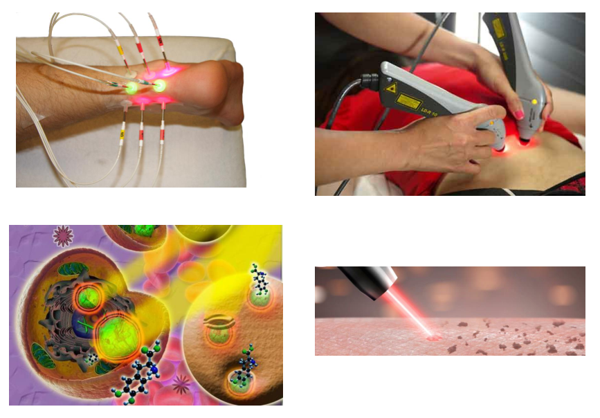
It was mentioned above that the laser can peel off the top layers of the skin. It is used in particular for tattoo removal. Beauty salons use a solid-state laser with a wavelength of 1064 nanometers to make tattoos.
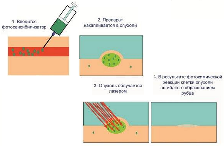
Another common application of lasers is photodynamic therapy, which is often used in the treatment of cancer. First, a photosensitizer is introduced into human tissue - a substance that accumulates in aggressive cancer cells. After that, the tumor - it is usually surrounded by healthy tissue - is exposed to a laser with a wavelength that falls within the absorption maximum of the photosensitizer. As a result, the radiation is absorbed only by cancer cells. Thus, we burn out the cancer without affecting healthy tissue.
The laser is used in medicine for imaging. For example, in optical tomography, it serves as a light source (see diagram). A superluminescent diode can also be used as a light source: it also emits due to stimulated scattering, but does not have this degree of coherence.
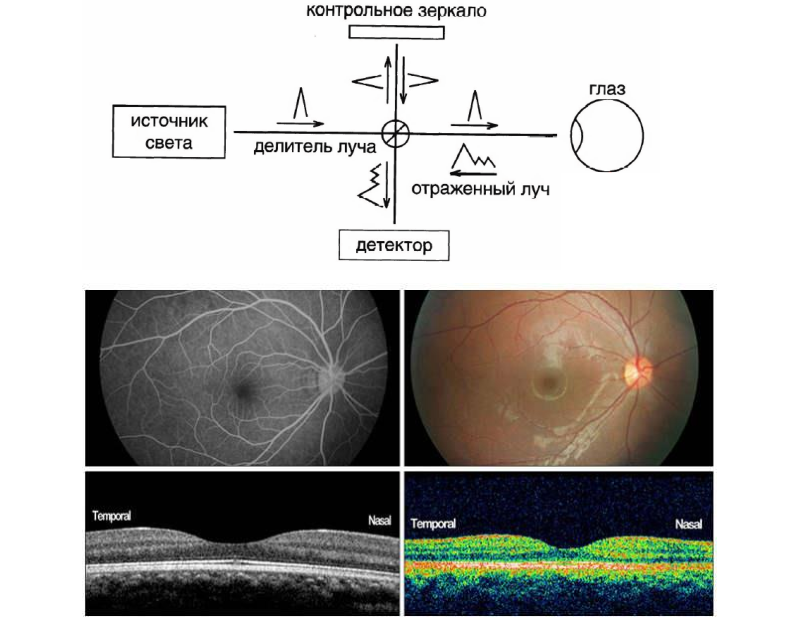
The light source is directed towards the beam splitter. Part of the radiation is reflected on the mirror, and the other is directed to the object, reflected from which both waves can interact with each other. If two coherent wavelengths interact with each other, interference occurs. And on the detector we register a set of interference fringes, after processing which we can get a picture of a tissue section.
An optical coherence tomograph, the principle of which is shown in the diagram, is available in all major cities. This technology allows you to build a three-dimensional picture of an object, in this case, the eyes. And the spatial resolution, where we can separate one pixel from another, can be a few microns. An analogue of this technology is ultrasound. Only for ultrasound, not optical radiation is used, but an ultrasonic wave. Ultrasound has a higher penetration depth, which cannot be said about accuracy: spatial resolution is measured in millimeters, not microns.
Why you need to combine methods
At Samara University, this approach was used to study skin and lung tissues with oncological formations. The photo on the left is a reconstructed 3D image of lung tissue. And on the right is a photograph of the area from which the signal was recorded.
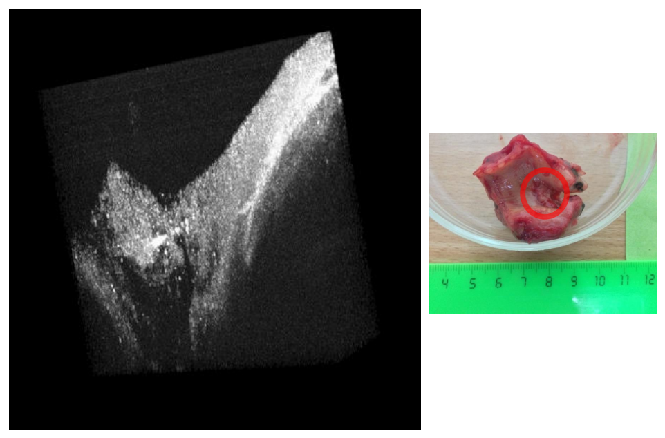
The picture on the left shows the difference between the structures. Black is air, no signal came from there. The sponge-like porous structure is healthy lung tissue. Moving to the right, you can see how layers are formed. They are denser and have a certain structure, which is characteristic of oncological neoplasms in the lung tissues. This is an example of squamous cell carcinoma removed as a result of an operation at the Samara Cancer Center.
The same approach was used to study skin tissue. It makes it easy to identify basal cell carcinoma, but other types of cancer are often similar to each other, and it becomes impossible to diagnose a specific type of disease. Therefore, optical research methods must be supplemented with spectral ones.
The following illustration shows a diagram of Raman (inelastic) light scattering, the so-called Raman scattering. Here we again observe the energy levels that we got acquainted with when considering stimulated scattering.
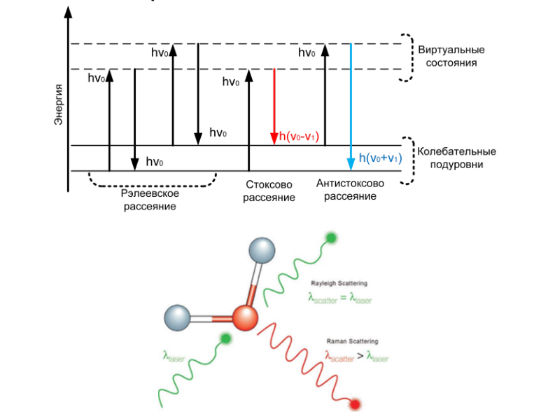
The picture shows how laser radiation excites vibrations in a molecule. Moreover, 99.999% of this radiation does not change the wavelength. But some part of the radiation after interacting with the molecule can change. This fraction of the energy change corresponds to the vibration of the bonds to which the laser radiation was directed.
As a result of Raman scattering of light, we get a set of bands, the position of which is tied to a specific vibration of our object. With this data, we can determine what fluctuations we have. In turn, the quantitative composition of these components is determined by the vibration intensity.
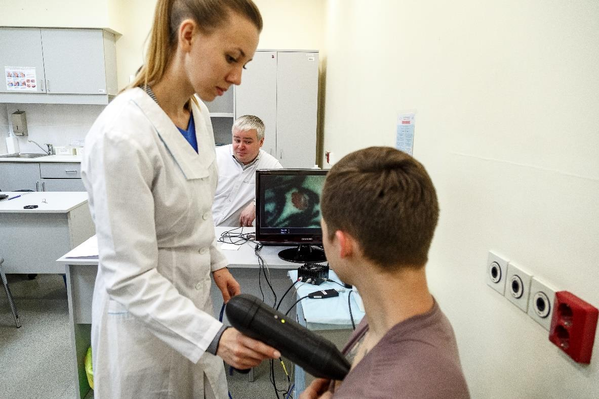
The photo shows the moment of research at the Samara Cancer Center. This is how a tissue sample is visualized using a dermatoscope developed there.
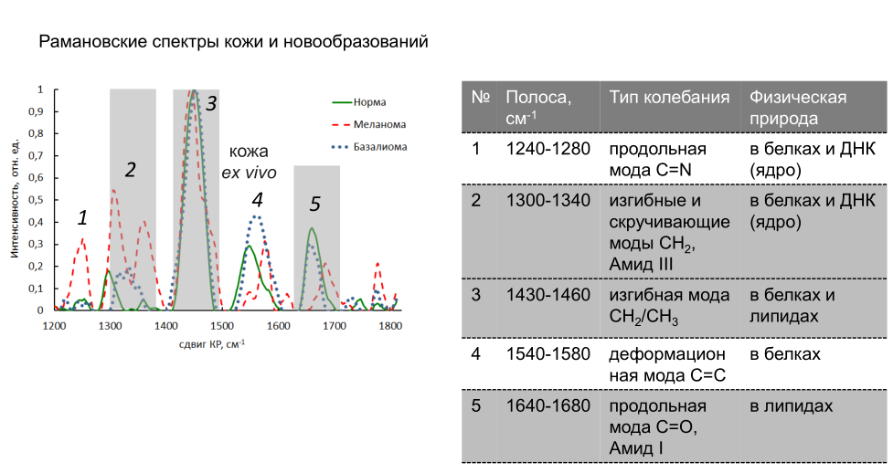
The next slide shows characteristic graphs of Raman spectra for skin and neoplasms. In certain bands of the spectrum, the intensity can increase or decrease. So, in lane 2, the intensity for malignant melanoma increases by 100%. And the change in the component composition in this area is responsible for the increase in this intensity. In particular, if we are talking about biochemical changes in tissue, then the ratio of DNA and RNA in the cell changes. The ratio of proteins to lipids in the tissue may also change.
A similar study was carried out for lung tissue. We see that it is possible to distinguish malignant tumors from benign ones. Also, various mathematical approaches can be used for data analysis - for example, regression models, which allow you to quickly find spectral differences in a large data set.
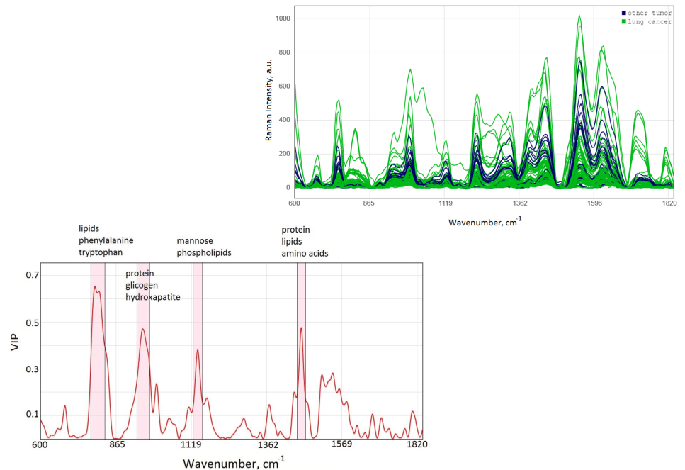
So, the study of a biological object using lasers and spectral technology allows you to get a huge set of data. To process them, one has to resort to mathematical methods, which, in turn, must be implemented on a computer using special software.
Let's sum up
Biophotonics provides ample opportunities for diagnosing the state of tissues in real time, allows for laser ablation - cleansing the upper layers of the skin. The laser scalpel is widely used in surgery. Also, when a laser is irradiated in the body, some processes can be accelerated, for example, the production of oxygen in blood vessels or some tissues. Or slow down if necessary.
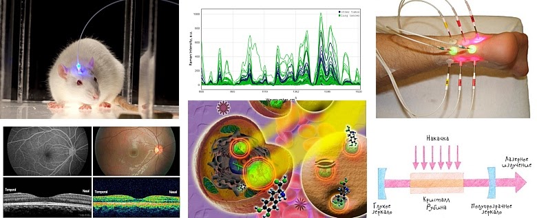
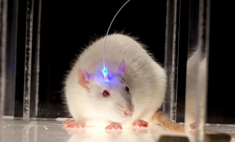
All optical technologies are used for non-invasive research - without direct contact of the instrument with the tissue. For more accurate research in different ranges, you can use several lasers at once. But these are not all possibilities. We did not mention such an interesting direction as optogenetics - the effect of laser or optical radiation on cognitive functions. Researchers target neurons in specific areas of the brain to try to improve mood, stimulate hormone production, and so on. While such experiments are being carried out on animals. The photo shows a mouse with an optical fiber implanted in its skull for appropriate research.
In connection with the current pandemic, it is worth noting that the aforementioned Raman spectroscopy is a technology that can be used to research viruses. Here again, an interdisciplinary approach: viruses are particles 20-200 nanometers in size, you need to catch them somehow. Viruses are contained in the blood, which moves through a certain capillary. Consequently, special nanotraps are installed in the capillary - nanostructures capable of trapping and capturing particles of a certain size. After the particles are captured, we carry out their irradiation with a laser and registration of Raman scattering - now we can say for sure what it is. The advantage of optical technologies in this case is that viruses are detected even at their minimum concentration.
***
In our opinion, we have listed most of the most interesting areas of laser application. Although they probably could have forgotten something. So, if someone throws in interesting facts in the comments, we will pop with pleasure.